
Hydration Effect of Geneo X Actions on Human Skin
Natural mechanisms of skin hydration
Skin hydration, referred to elevation of the water content, is essential in regulating its physical and mechanical properties of cutaneous tissue. Decreased hydration leads to two distinct conditions - dry skin and dehydrated skin. Dry skin is associated with natural deficiency in sebum production, which can be worsened with age or harsh skincare products. Dehydrated skin is caused by lack of water in its protective barrier due to sun exposure, harsh weather, or insufficient water drinking.
Epidermal and dermal hydration
The epidermis and dermis are the primary skin layers contributing to the natural skin hydration. The epidermis is divided into the outermost non-viable stratum corneum (SC) and the underlying viable layers. The dermis is rich in blood capillaries and consists of a superficial papillary layer and a thick, irregular reticular layer.
It was shown that therapeutic skin hydration mainly affects the stratum corneum, which forms the main barrier between the body and the environment. The SC is 10–15 μm thick and made up of dead, flattened corneocyte cells embedded in lipid matrix and filled with rigid keratin filaments, acting as a mechanical scaffold (Figure 1). The corneocytes are continually shed and replaced by the underlying layers, contributing to the skin's renewal and protection. The lipid matrix, composed of ceramides, fatty acids, and cholesterol, helps to seal in moisture. The hydrolipid film on the skin's surface and the intercellular lipid bilayer play a role in preventing water evaporation.1
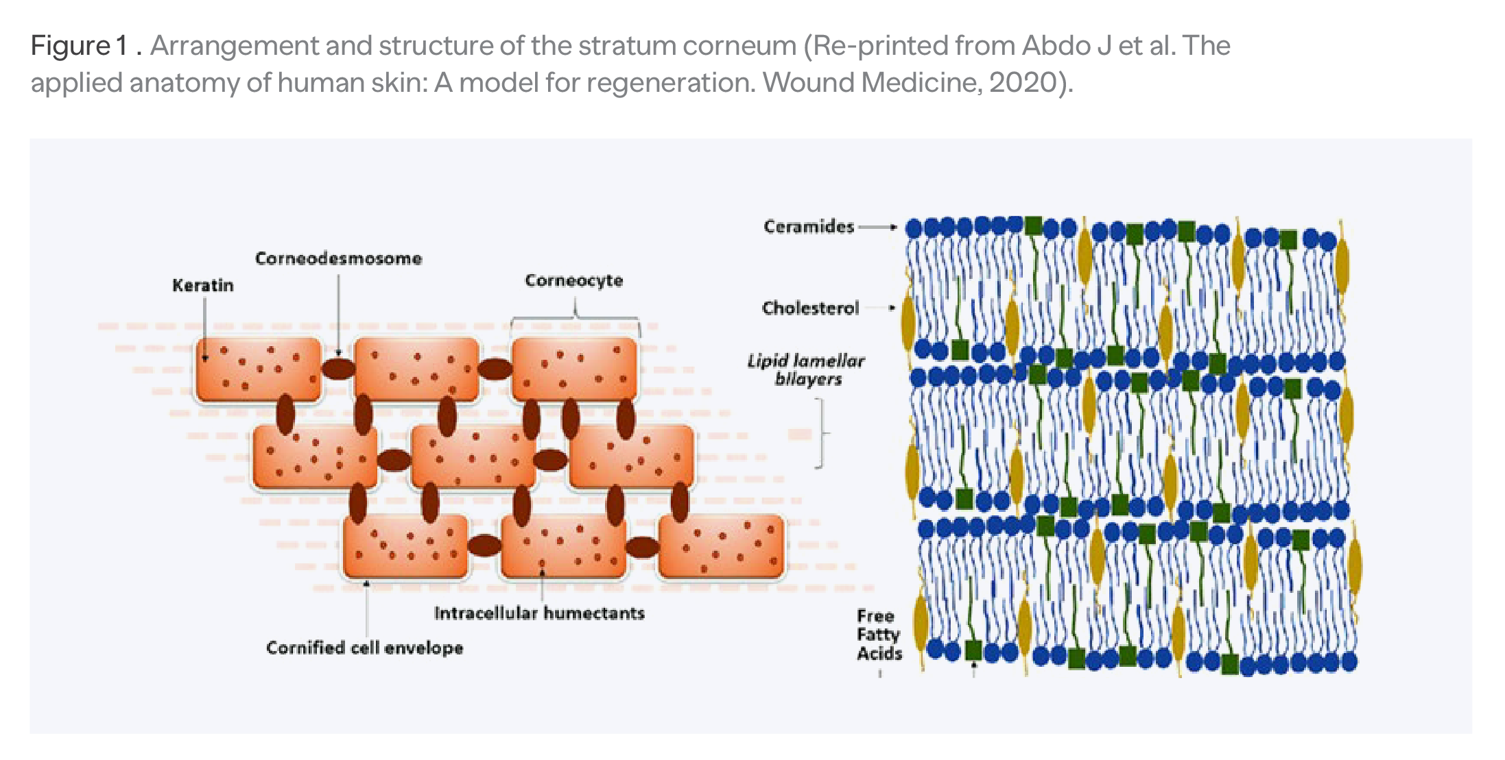
Figure 1. Arrangement and structure of the stratum corneum
(Re-printed from Abdo J et al. The applied anatomy of human skin: A model for regeneration. Wound Medicine, 2020).
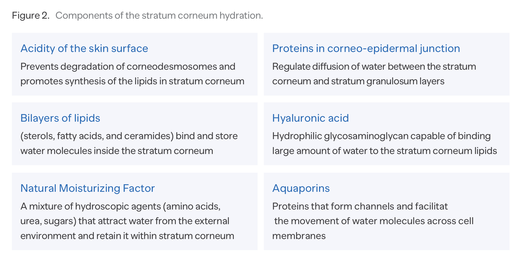
Physical properties of the skin cannot be warrantied without adequate SC hydration.2,3 Well-hydrated SC is characterized by increased keratin mobility, swelling of the corneocytes and the presence of intercellular aqueous inclusions (i.e., aqueous pools of varying sizes) in the SC.4 Figure 2 identifies 6 components controlling physiological hydration mechanism.
Studies have shown that SC hydration has a strong impact on its barrier properties. Altered barrier function is recognized as a significant part of the pathophysiology of inflammatory skin conditions and shows increase in trans-epidermal water loss to the external environment.5
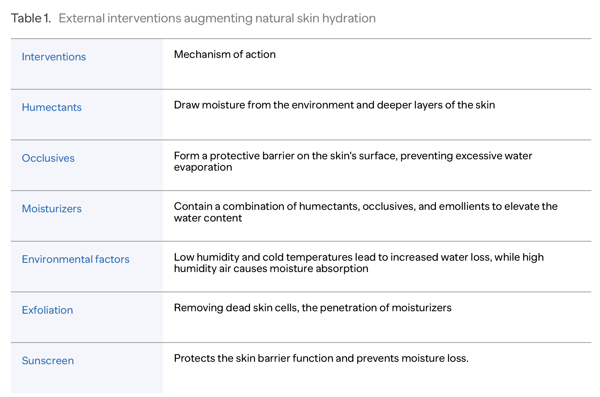
Various therapeutic interventions are widely used to elevate the skin hydration and augment its natural barrier function. (Table 1)
Opposite to epidermis, the dermis is hydrated mainly through internal mechanisms, including interstitial fluid and the spacing components. The fluid is generated from the flowing capillary blood, through the mechanism of the transcapillary exchange. Water-binding glycosaminoglycans (particular, hyaluronic acid) fill up the extracellular space, ensuring elasticity and firmness of the skin.6
Geneo X
The Geneo X device (Pollogen, Ltd) is a multi-technology platform, delivering a complex regenerative approach for the aging skin (Figure 3).
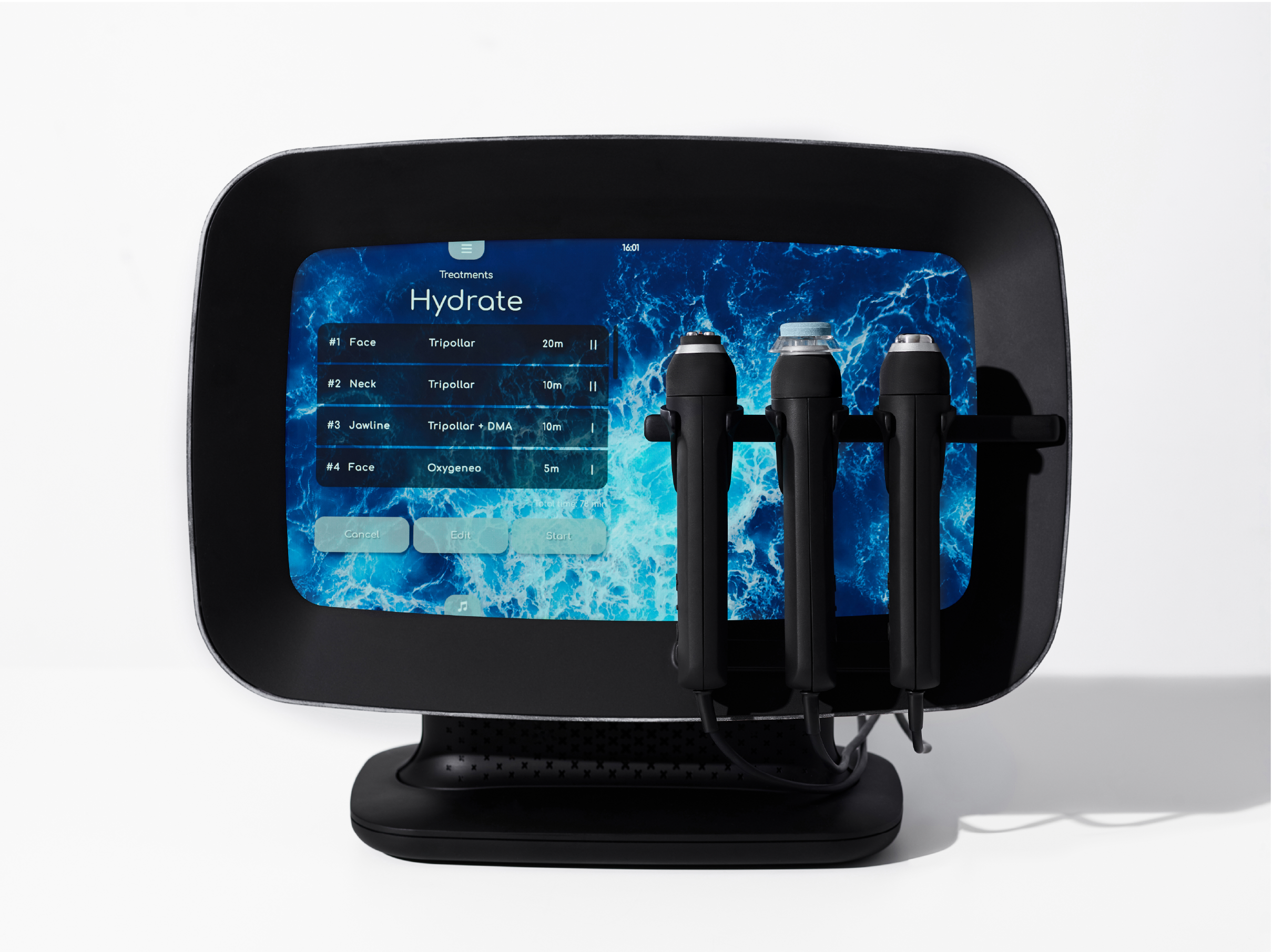
Figure 3. Geneo X device with multi-functional handpieces
providing external cutaneous oxygenation with OxygeneoTM, skin tightening with TriPollarTM radiofrequency, skin regeneration with electrical skin activation (ESATM), along with perfusion-stimulating massage and ultrasound-assisted absorption of the topical therapeutics.
Oxygeneo technology
Oxygeneo technology utilizes physiological mechanisms of the Bohr’s effect, according to which elevated concentration of carbon dioxide (CO2) decreases hemoglobin’s affinity for oxygen and eases its upload into cutaneous tissues.7 The technology implements Oxypod tablet containing citric acid and sodium bicarbonate. The chemical reaction occurring between these components generates a large amount of CO2 bubbles and water on the skin surface.
Abrasive Oxypod surface exfoliates the outmost skin, making the epidermal barrier permeable for CO2 diffusion.8 Triggered by increased CO2 concentration in the skin, cutaneous microcirculation delivers more O2-rich blood to the tissues, manifested by the rise in transcutaneous oxygen pressure.9
Ultrasound technology
The process is enhanced by the low-frequency non-focused ultrasound. The ultrasound vibrations further reduce the density of the stratum corneum, easing absorption of the topical active ingredients. The vibrations additionally result in a slight thermal effect and lead to increase in cutaneous microcirculation.
TriPollar RF
Oxygeneo is usually combined with non-ablative TriPollar radiofrequency (RF) treatment, which generates heat though utilizing the electrical resistance within the skin layers. Propagation of
the electrical current through the skin tissues oscillates the charged particles and generates the uniformed heat within 37-42°C range to dermal layers.
The resulting contraction of the existing collagen and synthesis of the new collagen causes immediate skin tightening and improved skin laxity lasting for months.10 Potekaev11 observed a significant reduction of static facial wrinkles and improved homogeneity of the facial skin color in 20 women aged 35-65 who received 8 weekly TriPollar treatments.
The findings were objectively justified with 3D scanning, ultrasonic micro-topography, and Doppler assessment of cutaneous micro-circulation.
Oxygeneo hydration studies
Recent clinical investigations evaluated facial skin moisture following Geneo X treatments. The investigations involved combination of Oxygeneo, TriPollar RF, and the ultrasound treatments in a group of healthy volunteers. The investigations were performed in the ISO-accredited and GMP & GLP certified QACS Labs (Athens, Greece).
Each participant (male and female, age range 20-50 years old) received a course of 6 Oxygeneo-and-ultrasound procedures once-a-week (with or without pre-procedural TriPollar RF) (Table 2). During the study period, participants abstained from hydrating cosmetic products, harsh cleansing soap, and exposure to intense sun or solarium.
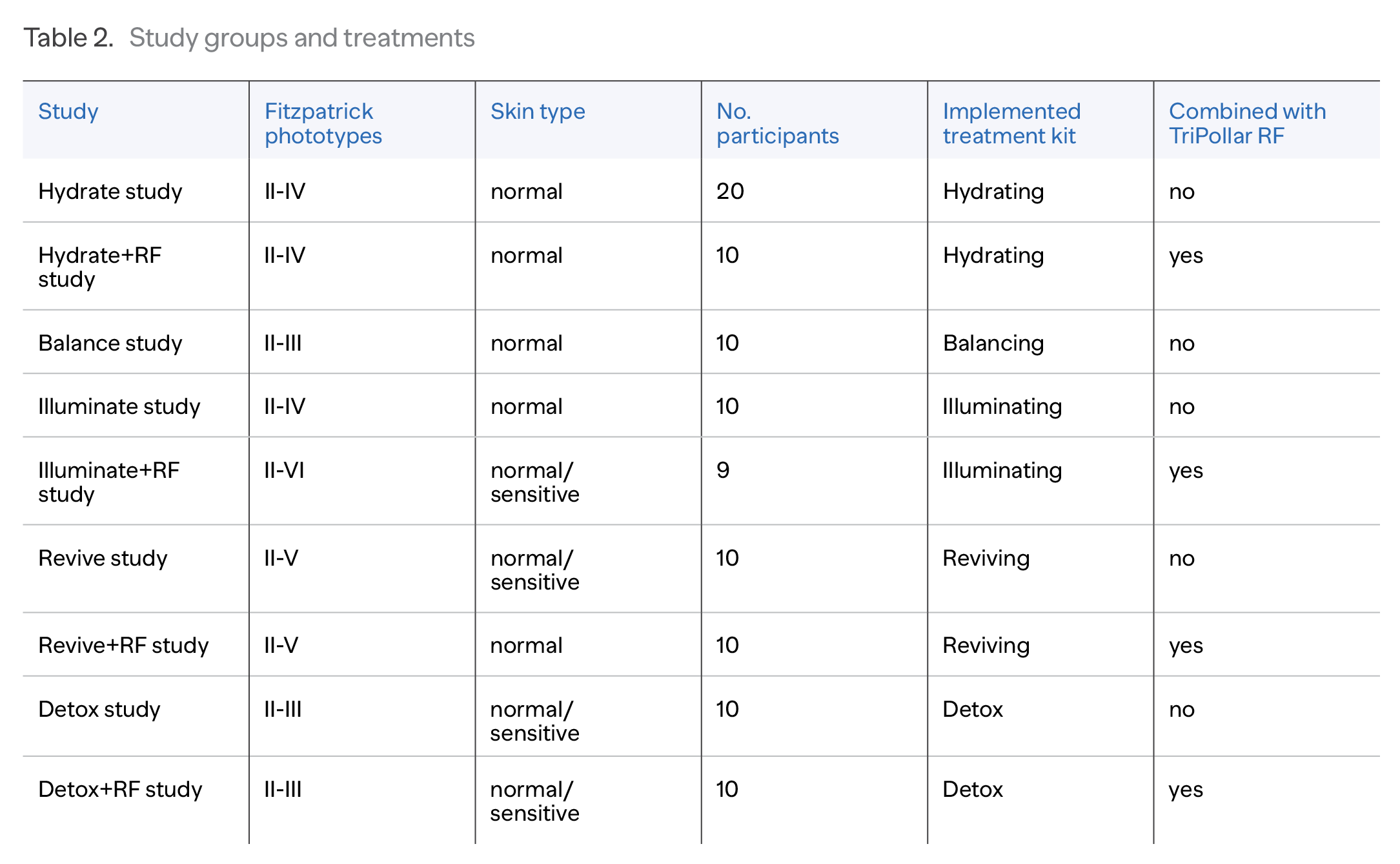
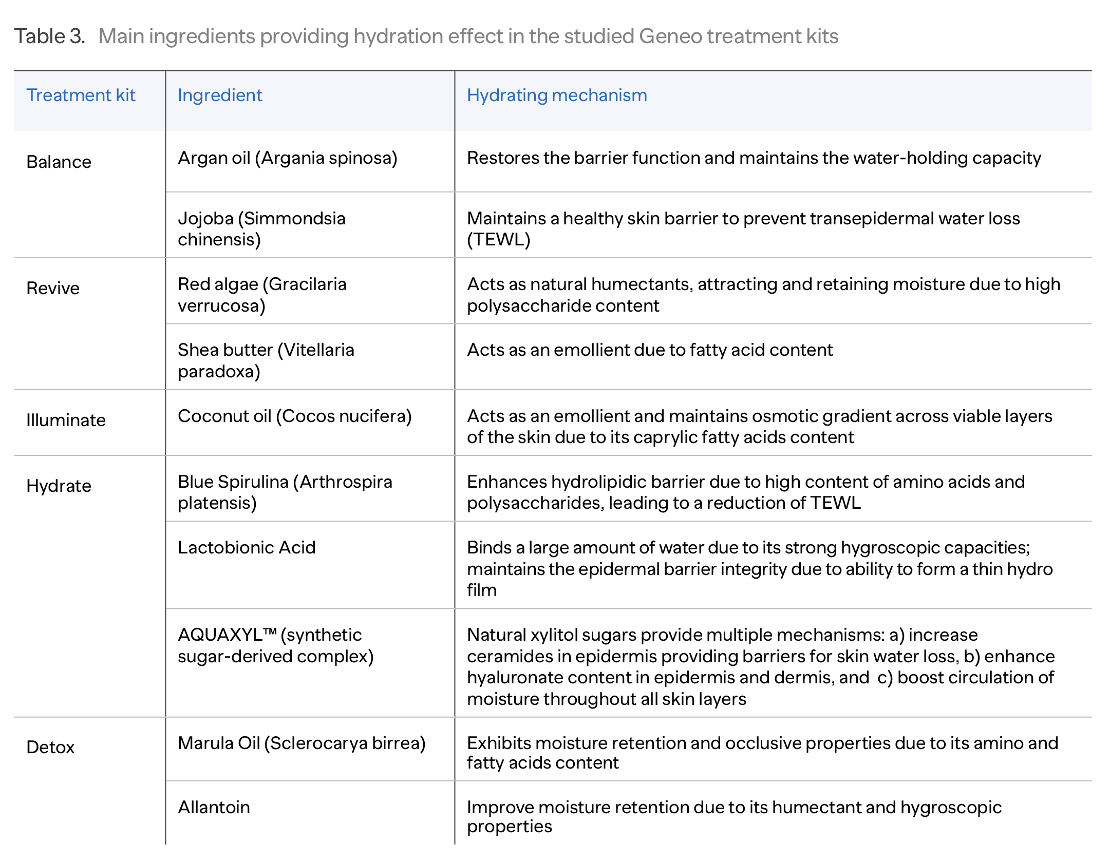
Oxygeneo procedures were performed using a unique set of non-allergenic natural ingredients piled in Hydrating, Balancing, Illuminating, Reviving, and Detox treatment kits. The constituents included in the tested Oxypods and serums exhibited various natural mechanisms to attract and retain a significant amount of water in the skin (Table 3).
End-point measures
The skin was clinically and instrumentally assessed at baseline (T0), mid-course (T3), and 2 weeks after the last treatment (T8). Dermal hydration was assessed at different levels using multi-probe measuring device (Courage+Khazaka electronic GmbH, Köln, Germany).
1. Hydration of the stratum corneum was measured with the Corneometer® CM 825 probe. Corneometer measures the capacitive resistance of the stratum corneum, reflecting its degree of hydration. Skin capacitance dependents on the water content and changes as the degree of hydration increases.
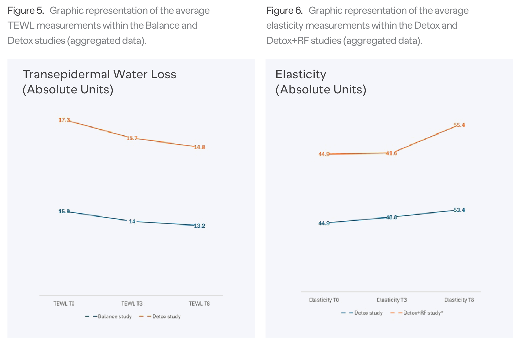
2. The ability of the skin barrier to retain moisture was assessed with Tewameter® TM 300 probe, which is based on diffusion measurement. The weakening of the barrier is reflected by increase in transepidermal water loss (TEWL), induced by inflammatory or environmental factors. Reduced TEWL demonstrates a strengthening of the skin barrier, characterized by an increased number of corneocytes or better organization of the lipid matrix.
3. Viscoelastic properties of the skin were measured with Cutometer® MPA 580, which measures the elasticity of the upper skin layers by applying a vacuum that mechanically deforms the skin. Elasticity is decreased as the skin dehydrates at viable epidermis depths.
Obtained results were statistically analyzed in comparison to baseline values using the t-test.
Results
Table 4 shows the average values of the skin hydration parameters collected in the studies.
The skin hydration measurements appear to be higher and significantly different at T3 and T8 compared to the baseline T0 (p<0.005). A similar increase can be seen in viscoelastic characteristics of the skin during and two weeks after the treatment course. The data also showed that TEWL measurements decreased within the study period, which indicated strengthening of the skin barrier after the treatments. (Figures 4-6)
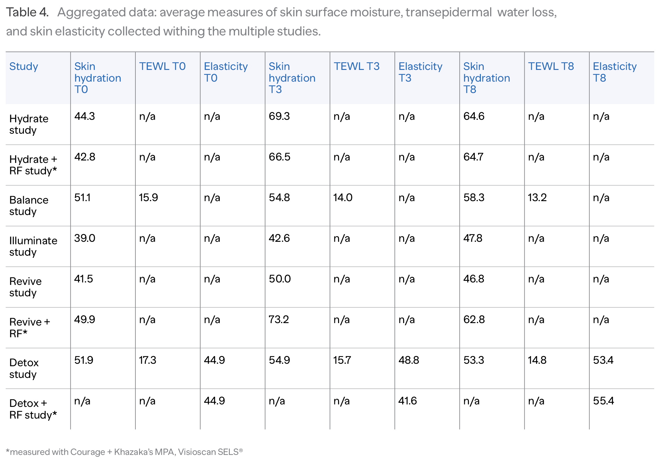
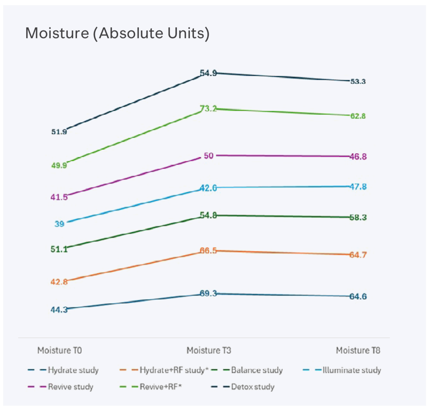
Figure 4. Graphical representation of the average skin moisture measurements within the Hydrate, Balance, Illuminate, Revive, and Detox studies and the studies combining TriPollar RF (aggregated data).
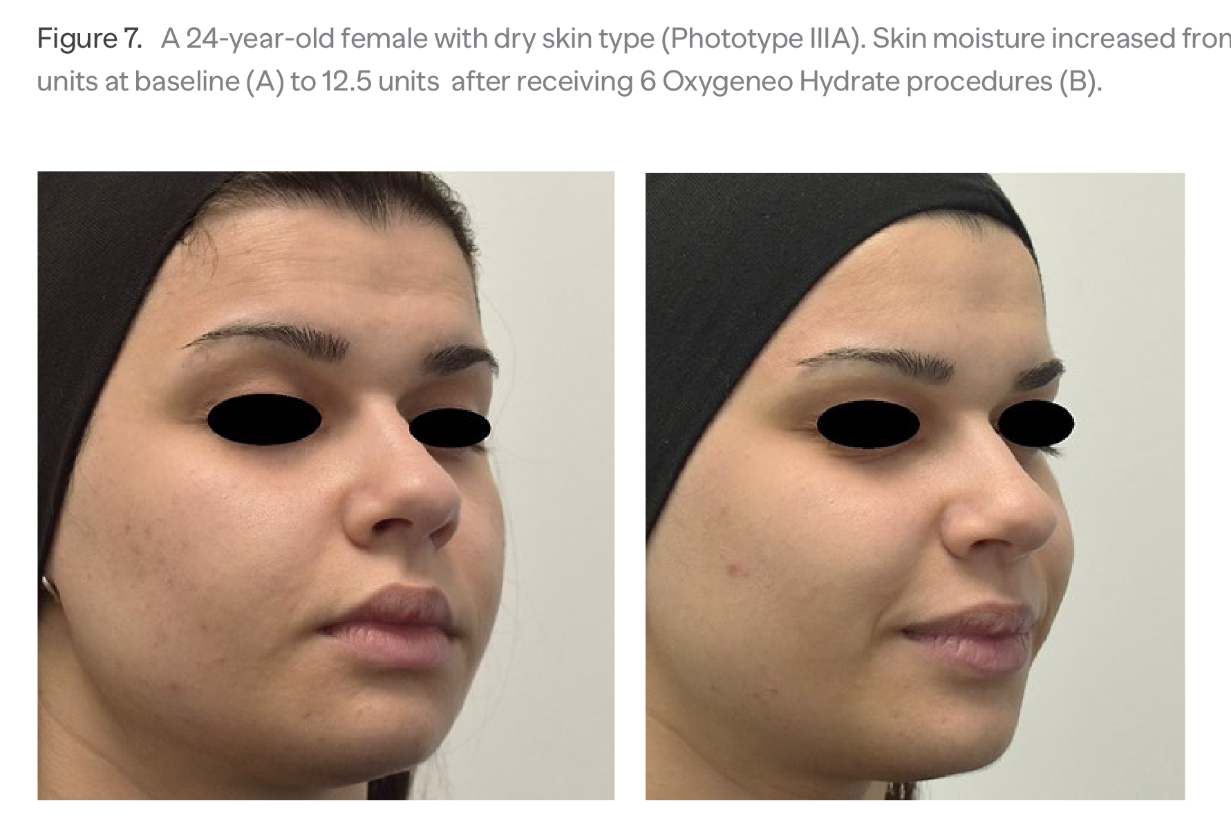
The results demonstrated increased skin hydration level following the course of Geneo procedures, which combined Oxygeneo, ultrasound-induced application of the hydrating serum, and TriPollar RF treatments. The increase was maintained 2 weeks after completing the treatment course. In addition to the objective measurements, a substantial percentage of the participants expressed their satisfaction with instant skin hydration after each treatment and with skin hydration after completing the full treatment course (data is not included in this report). (Figure 7)
Discussion
Each technology implemented in Geneo X provides, to certain extent, augmentation of skin hydration and beneficial stimulation of cutaneous microcirculation.
Mechanisms of skin hydration enhancement
Studies reported significant improvement in skin quality following therapeutic increase of CO2 level in the epidermis.12,13 Enhanced hydration of the stratum corneum was attributed to the reduction of skin erythema, flaking, and crusting. It was stipulated that therapeutic mechanism of Oxygeneo may be similarly associated with the hydration changes in SC. We proposed that pertaining mechanism is likely to have a dual nature (Figure 8):
1. Exogeneous cutaneous hydration is provided by the water abundantly originated from the chemical reaction between the components in Oxygeneo tablet. With the tablet exfoliating the skin surface, the water is easily absorbed by the stratum corneum.
2. Endogenous cutaneous hydration is supplied though the liquid components of the blood perfusing the dermis. Vasodilation caused by the CO2 elevation raises capillary hydrostatic pressure to the level at which the liquid from capillaries escapes, so the fluid volume within the interstitial space increases.
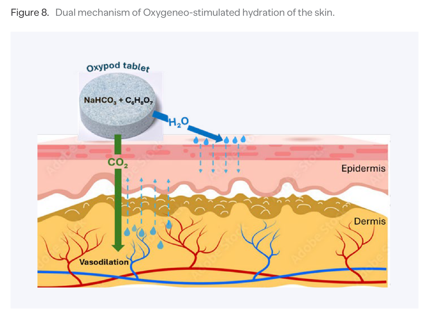
Figure 8. Dual mechanism of Oxygeneo-stimulated hydration of the skin.
Procedural hydration is enforced with absorption of the Oxypod ingredients, which follow intradermal path created by the exfoliation. Ingredients described previously, increase hydration content, lower TEWL, and overall repair the skin barrier. Its protective function is further restored by the abrasive surface of the Oxypod tablet. Exfoliation facilitates the cell turnover, with the new cells enhancing structural integrity of stratum corneum by establishing new desmosome cohesion.14
Furthermore, hydration effect of radiofrequency-generated heat was previously demonstrated in the study of Kruglikov.15 The skin reacts to RF-generated heat by activation of fibroblasts and increased concentration of hyaluronan and other glycosaminoglycans, known for their ability to bind water and helping to keep the skin hydrated. Expansion of the papillary dermis due to edema and vascular congestion was observed in the study of Alvarez16 after dermal application of radiofrequency.
In the study of 62 healthy women receiving facial treatment with bipolar RF device, Palmieri17 revealed improvement of total water content, elasticity, and the microvascular restoration. Stochaj18 demonstrated sustainable epidermal hydration in the facial skin of adult females 4 months after bi-polar RF treatments.
Mechanisms of microcirculation augmentation
Skin microcirculation refers to the network of tiny blood vessels in the dermis (arterioles, venules, and capillaries) organized in two parallel plexuses with capillary loops extending perpendicularly from the superficial plexus. (Figure 9) It plays a vital role in supplying epidermis and dermis with water and nutrients. Skin blood flow is highly adaptive and quickly responds to various stimuli, like temperature changes. Additionally, it can be enhanced with Oxygeneo procedure. Trans-cutaneous diffusion of CO2 reduces tissue pH and inhibits contractility of the smooth muscles in the vessel walls. Resulting immediate vasodilatation opens nonfunctioning skin capillaries to blood flow and facilitates the capacity of hemoglobin to release O2. In the study of 12 patients, Seidel9 demonstrated statistically significant increase of transcutaneous O2 tension (TcPO2) from baseline 51.56±3.53 mmHg to 62.85±2.64 mmHg after Oxygeneo facial treatment (p<0.05).
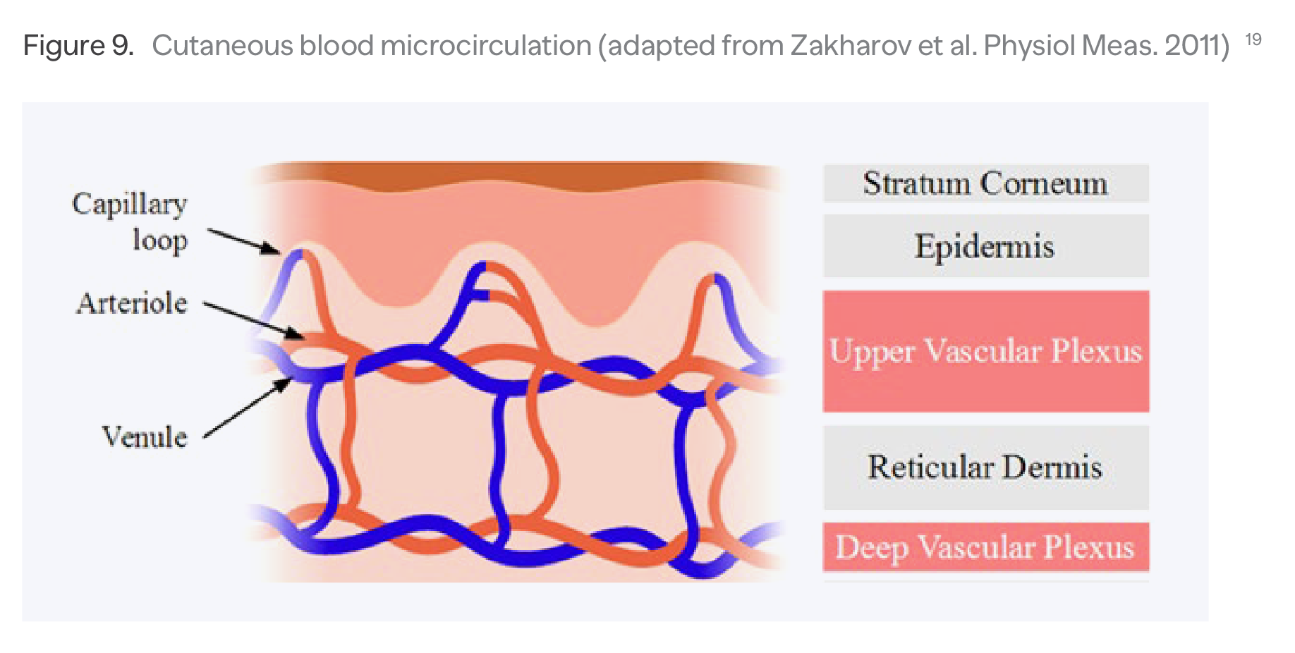
Figure 9. Cutaneous blood microcirculation (adapted from Zakharov et al. Physiol Meas. 2011)19
Levenberg10 measured cutaneous blood perfusion following Oxygeneo procedures in a group
of 11 healthy male and female subjects. Assessed with the laser Doppler, an average cutaneous microcirculatory blood perfusion increased by 16 units immediately after the treatment and maintained 10 units above the baseline 15 minutes after treatment. The accompanying transcutaneous oximetry revealed sustained increase of the skin oxygen from the pre-treatment 37.8±1.6 to 73.0±3.0 in 15 minutes after Oxygeneo.
Besides promoting adequate oxygenation, repeated elevation of cutaneous CO2 induces local angiogenesis, or physiological formation of new blood vessels. Irie20 reported elevation of plasma vascular endothelial growth factor and increased number of endothelial progenitor cells following prolonged skin exposure to CO2. Added angiography revealed the formation of collateral vessels
and increase of the capillary densities. A clinical study of Leibaschoff21 demonstrated that the transcutaneous application of a topical CO2 led to an increase in vertical and horizontal capillary density and significant improvements in cutaneous microcirculation.
It can be further facilitated with the thermal effect generated by Geneo X’s TriPollar RF. Radiofrequency energy produces uniform heat at controlled depth to dermal layers, resulting in
the immediate skin tightening.22 The heat indices a relaxing effect on the smooth vascular muscles resulted in vasodilation of dermal arterioles and recruitment of capillaries.23 The Doppler flowmetry study24 demonstrated increased blood flow in the microcirculation as much as 15-20 times. With cutaneous vasodilation and increased flow rate, skin perfusion is re-arranged to bring more blood and interstitial fluid to the skin surface.25
In addition, Geneo X’s ultrasound complements microcirculation by generating a mild rise in skin temperature. The thermal effect comes from the collision of skin and ultrasonic waves, which create tissue friction and successfully generates the tissue heating. The heat dilates superficial capillaries and activates the capillary circulation.26 Augmented blood flow is expressed by appearance of skin erythema (Figure 10).
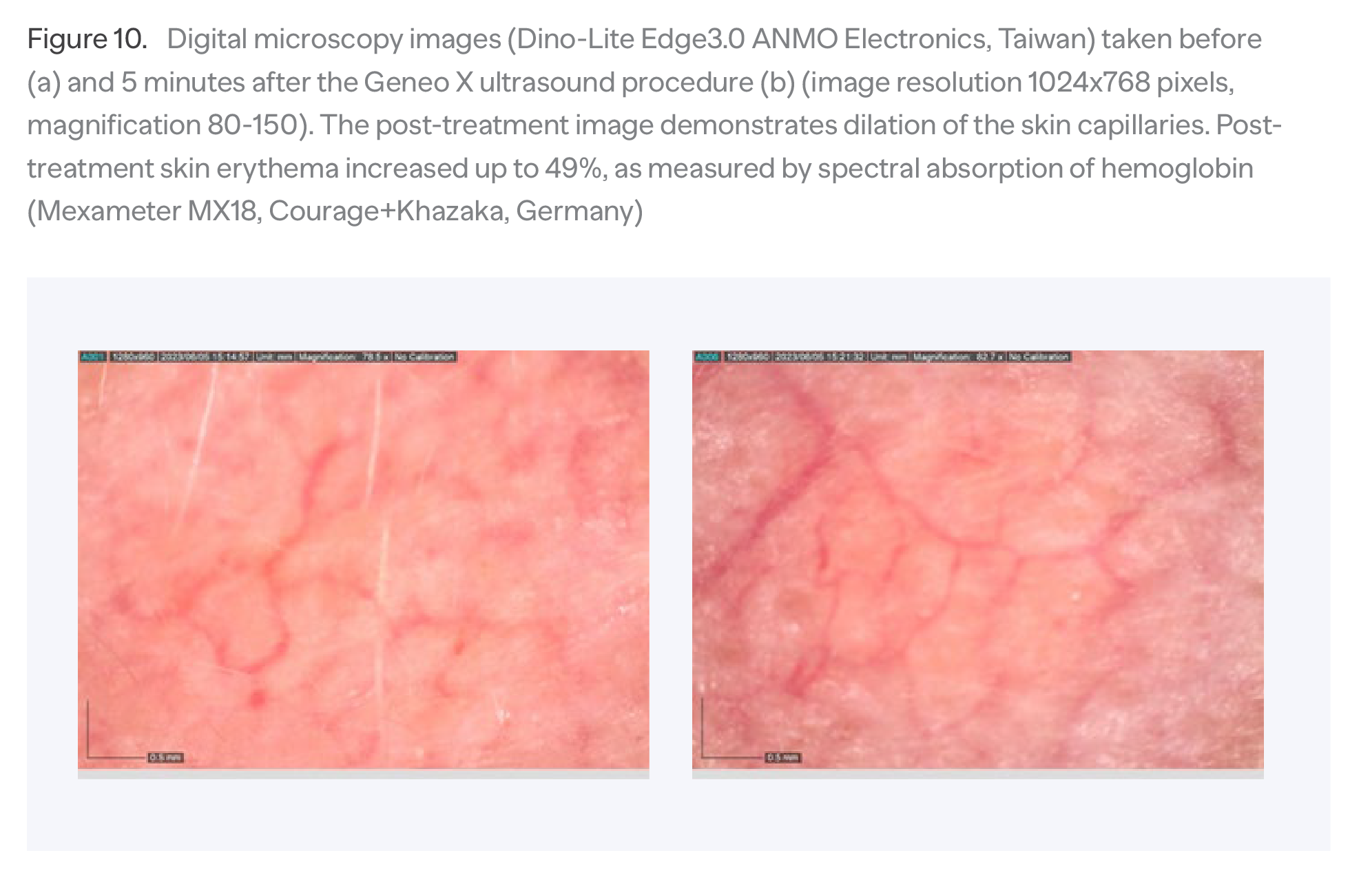
Figure 10. Digital microscopy images (Dino-Lite Edge3.0 ANMO Electronics, Taiwan) taken before (a) and 5 minutes after the Geneo X ultrasound procedure (b) (image resolution 1024x768 pixels,
magnification 80-150). The post-treatment image demonstrates dilation of the skin capillaries. Post-treatment skin erythema increased up to 49%, as measured by spectral absorption of hemoglobin (Mexameter MX18, Courage+Khazaka, Germany)
Rational to combine Geneo X technologies with aesthetic energy-based treatments
The correct strategy for preparing the skin before the energy-based treatments is the best possible way to avoid adverse effects and ensure optimum results.
Multi-technological Geneo X is capable to address physiological processes of increasing skin hydration, restoring skin barrier, augmenting cutaneous microcirculation, and raising the basal tissue oxygenation. Therefore, it seems rational to combine these clinical advantages with various forms of therapeutic energy-based devices. In the treatments addressing cosmetic concerns, Geneo X can be utilized either as skin preparation or post-treatment skin rehabilitation.
Ablative and non-ablative lasers
a) For the lasers targeting water chromophore, absorption of the laser energy is proportional to the water content in epidermis27,
b) Well-hydrated skin provides a higher ablation volume and more intense volumetric vaporization compared to dehydrated skin28,
c) Pre-treatment skin exfoliation lowers loss of energy due to the refraction and scattering of the light in the stratum corneum layer29,
d) Pre-treatment elevation of the skin temperature lowers the electrical impedance properties of the skin barrier30,
e) Moisture-retention applied to the laser-treated skin results in faster recovery of cutaneous erythema and faster functional recovery of the skin barrier31,
f) Sufficient skin oxygenation is essential for the healing mechanism of the thermal wounds: fibroblast proliferation, formation of collagen matrix, wound granulation, and reepithelization.32
Intense pulsed light (IPL)
a) Pre-treatment induction of skin hydration is a practical solution for post-IPL depletion of skin moisture33,
b) Oxygeneo activates intracellular activity and improves skin nourishment – all to prevent the stress generated in the skin by exposure to intensive light energy.
c) Moisturized stratum corneum benefits its optical properties resulting in less light scattering at the skin surface and more light penetraation into the deeper skin layers.34
Light-emitting diodes (LED)
a) Complementary interaction of LED and Oxygeneo contributes to cutaneous regeneration through synergistic augmentation of blood flow and enhanced production of mitochondrial ATP35,
b) Harmful synergy of LED and Oxygeneo on the anerobic acne bacteria – through increased skin oxygenation and LED-stimulated damage of bacterial cell membranes and essential cellular components.36
Focused ultrasound (HIFU, MFU)
a) Adequate hydration supports the skin's natural ability to produce collagen, a key factor in focused ultrasound effectiveness,
b) Well-hydrated skin can better withstand the heat generated during the procedure, minimizing the risk of irritation or discomfort37,
c) Combining HIFU with RF demonstrated an improved post-treatment skin moisturization.38
Conclusion
Reviews of the literature and experimental data demonstrated the ability of the Geneo X technologies to augment skin hydration and boost cutaneous microcirculation. That provides rational for its implementation in the therapeutic combination with the aesthetic energy-based treatments.
References
1. Chao PH, Lu HH, Hung CT, Nicoll SB, Bulinski JC. Effects of applied DC electric field on ligament fibroblast migration and wound healing. Connect Tissue Res. 2007;48(4):188-197.
2. Jennings J, Chen D, Feldman D. Transcriptional response of dermal fibroblasts in direct current electric fields. Bioelectromagnetics. 2008;29(5):394-405.
3. Galan, Edgar & Bayat, Ardeshir. (2018). Alternating current electrical stimulation effects in human dermal fibroblasts. MACE PGR Conference University of Manchester, UK, March 26, 2018
4. Lee YI, Choi S, Roh WS, Lee JH, Kim TG. Cellular Senescence and Inflammaging in the Skin Microenvironment. Int J Mol Sci. 2021;22(8):3849.
5. Campisi J. The role of cellular senescence in skin aging. J Investig Dermatol Symp Proc. 1998;3(1):1-5.
6. Bhatia-Ney 2016 Cellular Senescence as the Causal Nexus of Aging
7. Ho CY, Dreesen O. Faces of cellular senescence in skin aging. Mech Ageing Dev.021;198:111525.
8. Bourdens M, Jeanson Y, Taurand M, Juin N, Carrière A, Clément F, Casteilla L, Bulteau AL, Planat-Bénard V. Short exposure to cold atmospheric plasma induces senescence in human skin fibroblasts and adipose mesenchymal stromal cells. Sci Rep. 2019 Jun 17;9(1):8671.
9. McCart EA, Thangapazham RL, Lombardini ED, Mog SR, Panganiban RAM, Dickson KM, Mansur RA, Nagy V, Kim SY, Selwyn R, Landauer MR, Darling TN, Day RM. Accelerated senescence in skin in a murine model of radiation-induced multi-organ injury. J Radiat Res. 2017 Sep 1;58(5):636-646.
10. Li G, Zhu Q, Wang B, et al. Rejuvenation of Senescent Bone Marrow Mesenchymal Stromal Cells by Pulsed Triboelectric Stimulation. Adv Sci (Weinh). 2021;8(18):e2100964.
11. Galan, Edgar & Bayat, Ardeshir. (2018). Alternating current electrical stimulation effects in human dermal fibroblasts. MACE PGR Conference University of Manchester, UK, March 26, 2018
12. Ho CY, Dreesen O. Faces of cellular senescence in skin aging. Mech Ageing Dev. 2021;198:111525.
13. Wlaschek, M.; Maity, P.; Makrantonaki, E.; Scharffetter-Kochanek, K. Connective tissue and fibroblast senescence in skin aging. J. Investig. Dermatol. 2021, 141, 985–992.
14. Chao PH, Lu HH, Hung CT, Nicoll SB, Bulinski JC. Effects of applied DC electric field on ligament fibroblast migration and wound healing. Connect Tissue Res. 2007;48(4):188-197.
15.
16. Jennings J, Chen D, Feldman D. Transcriptional response of dermal fibroblasts in direct current electric fields. Bioelectromagnetics. 2008;29(5):394-405.
17. Rouabhia M, Park H, Meng S, Derbali H, Zhang Z. Electrical stimulation promotes wound healing by enhancing dermal fibroblast activity and promoting myofibroblast transdifferentiation. PLoS One. 2013;8(8):e71660.